Stereotactic breast biopsy helps locate abnormalities
While physicals, mammography and other exams can often detect lumps in the breast, they can’t determine whether the growth is benign or cancerous. That’s why a procedure called stereotactic breast biopsy is often called upon.
Stereotactic breast biopsy is used when a small growth or an area of calcifications is seen on a mammogram. Stereotactic biopsy is an alternative to surgical biopsy. It’s a less invasive way to obtain the tissue samples needed for diagnosis, and the procedure requires less recovery time than a surgical biopsy. Dr. Stephen Manier, a radiologist with OSF HealthCare, says about 80% of the calcifications he biopsies are benign.
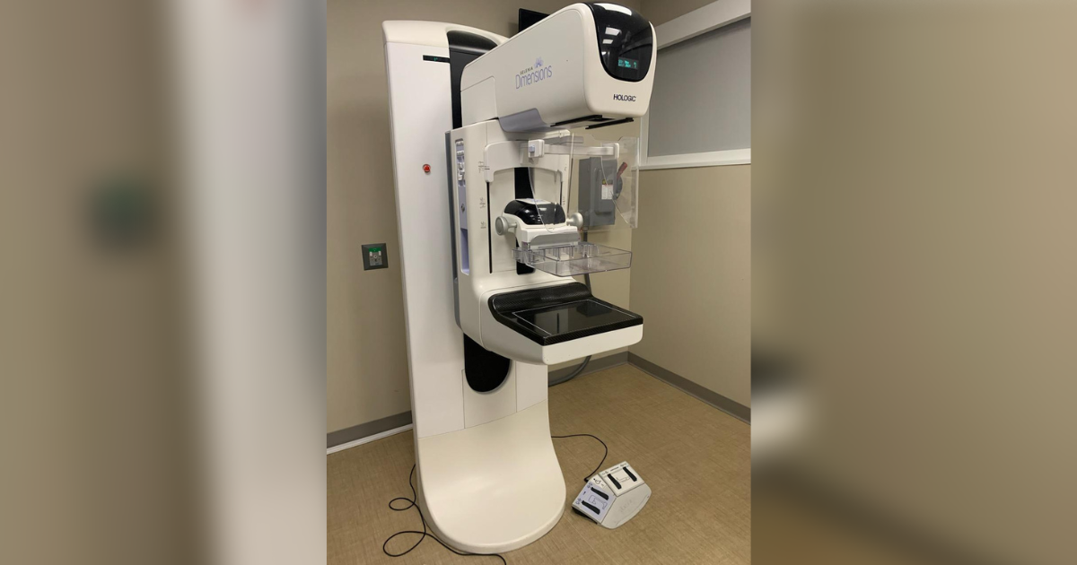
“In the past stereotactic biopsies were done with a special table where the patient’s laying on their stomach,” says Dr. Manier. “Those tables were usually only in places where they did a lot of those like big cities because they’re very expensive and you really can’t use those tables for a regular mammogram. So as a result of that, it’s hard to get a stereotactic biopsy without traveling. But in in the last several years, there’s another technique where you can actually do an add-on computer to a regular mammogram unit. And that’s what we do. That’s available in almost all hospitals now.”
Your physician may recommend a stereotactic biopsy if a mammogram finds any of the following:
- A suspicious lump
- Abnormal changes in breast tissue
- Irregularities in breast structure
- Changes in an area where surgery has taken place
“Most of the time these biopsies are done for breast calcifications,” says Dr. Manier. “The majority of the time, and it’s difficult to get a biopsy that way using anything else. Calcifications are not usually seen on ultrasound or MRI with breast issues. So doing a regular mammographic technique is really important.”
During a stereotactic biopsy a special mammography machine uses an x-ray technique to help guide the radiologist’s biopsy equipment to the site of the imaging abnormality. The radiologist uses images of the breast tissue to take a sample of tissue for testing, which is sent to a pathologist.
“There is compression on the breast. It’s similar to getting a mammogram,” says Dr. Manier. “Usually it’s not severe compression, so they’re not in pain, but it’s going to be firm. And it takes about 20 to 30 minutes to complete the test.”
According to the American Cancer Society, breast cancer is the most common cancer in women in the United States, except for skin cancers. It is about 30% (or 1 in 3) of all new female cancers each year. In 2022, about 287,850 new cases of invasive breast cancer will be diagnosed in women. That’s why the best way to treat cancer is to catch it at the earliest stage possible, and that’s where stereotactic biopsy can help.
“It’s a lot to go through for the patient,” says Dr. Manier. “But what happens is it’s very rewarding when you find that early breast cancer. You may or may not see a soft tissue density like a regular cancer often is seen, it’s often just calcifications but it can be an early tumor. And usually when you find things early, that’s a really good sign that you can take care of them. There’s a very high rate of cure when you find things early like that.” For more information, visit OSF HealthCare.
This article originally appeared on OSF Healthcare.